Calcaneal Insufficiency Fracture after Ipsilateral Total Knee Arthroplasty
Article information
Abstract
Insufficiency fracture of the calcaneus is a rare entity. In the absence of trauma, evaluating a painful ankle in an elderly patient can be difficult and also it might be overlook the insufficiency fracture. We experienced a case of insufficiency calcaneus fracture that occurred after ipsilateral total knee arthroplasty. Here, we report our case with a review of literatures.
INTRODUCTION
Insufficiency fracture is occurred by repetitive normal stress without significant abnormal force in the area of elastic resistance deficiency.[1] It is known to occur mainly in elderly female patients with osteoporosis.[2] In particular, following hip or knee joint arthroplasty, it occurs very rarely.[3] Asymptomatic cases of insufficiency fracture may be easily overlooked.[1] We describe one case of calcaneal insufficiency fracture following ipsilateral total knee arthroplasty with a review of literatures.
CASE
An 80-year-old woman underwent total knee joint arthroplasty due to the degenerative arthritis of the left knee joint. She did not have any history of trauma or coexisting medical illnesses. Her body weight was 45 kg, height was 153 cm and body mass index (BMI) was 19.34. She had been prescribed sodium alendronate and cholecalciferol composite agent (Fosamax plus D®; Merck & CO, Inc., West Port, PA, USA) for 1 year at local medical clinic. At a preoperative work-up, the patient received dual energy X-ray absorptiometry (DXA); this showed that the T-score of the femoral neck and lumbar spine were -3.6 and -3.1, respectively. Hematological investigations were within normal limits. A 5 days after operation, the crutch walking was started. The patient was discharged on 1 week after operation without significant complication. On postoperative month 3, the patient visited the outpatient clinic of the clinical department due to the presence of pain in the left heel. The patient told us that there was no trauma. On examination, there were edema and tenderness on the lateral and superior side of the left calcaneus. A compression test was also performed for the calcaneus; this also showed positive findings. There were no findings that are suggestive of burning sensation or erythema. On lateral and axial view of a plain radiography of the posterior region of the calcaneus, radiopaque lesions were shown as an irregular line that is parallel with the posterior joint of the subtalar joint (Fig. 1). The patient was informed of the possibility of insufficiency fracture and then prescribed with anti-inflammatory drugs. In addition, the patient was advised to limit a weight bearing during a gait. One month later, the patient visited the outpatient clinic again, presenting with a persistent presence of edema and pain again. A compression test was also performed for the calcaneus; this also showed positive findings. Radiopaque lesions that had been seen on lateral view of the diaphysis of calcaneus on plain radiography were found to be a more clear line (Fig. 2). Therefore, the patient underwent magnetic resonance imaging (MRI) scanning. On T2-weighted MRI scans, two linear lesions in the diaphysis of the calcaneus, which had not been seen on radiography, and they were perpendicular to the bone trabeculae. Thus, low-intensity signals were found. In the adjacent areas, there were high-signal intensity lesions that are suggestive of bone edema (Fig. 3). The patient was diagnosed with insufficiency fracture of calcaneus. To limit a weight-bearing effect, the patient underwent ankle-foot cast for approximately three months. Following the manifestation of symptoms, the patient achieved an improvement in them at a 2-month final follow-up. On plain radiography, the patient had a loss of radiopaque lesions. Moreover, the patient could also perform a normal gait.

Lateral and axial plain radiographs of the left calcaneus demonstrating radio-opaque irregular line (white arrow) on calcaneal body.
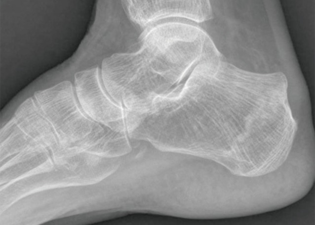
Lateral plain radiographs of the left calcaneus showing clearer radio-opaque line, being parallel to posterior facet of the subtalar joint compared to previous radiograph.
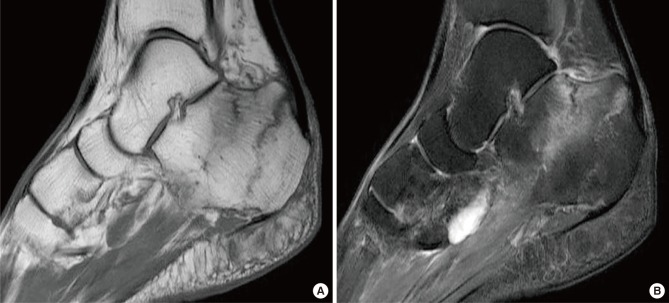
Sagittal T1-weighted image demonstrating two parallel lines with a low signal intensity (A), and T2-weighted image showing high signal intensity in the corresponding lesions (B).
DISCUSSION
Stress fracture is a term that is referred to as a fatigue one or an insufficiency one. It is a microfracture that occurs as a result of the abnormal stress to the normal bone, but an insufficiency one does when the normal loading of the daily living activity to the deficient bony elastic resistance.[14]
Insufficiency fracture is known to occur in mainly elderly female patients in association with osteoporosis, rheumatoid arthritis, hyperparathyroidism, diabetes mellitus, radiotherapy or steroid use.[5] It mainly affects pelvic ring, sacrum, tibia and the neck or head of the femur. Although rare, it may also occur in the talus, fibula and calcaneus.[3] There are also reports that insufficiency fracture occurred after total knee arthroplasty. Most cases in this series occur around the knee joint. Although extremely rare, however, it may also occur in the femoral neck or calcaneus.[67]
Still, little is known about the pathophysiologic mechanisms underlying the occurrence of insufficiency fracture in the ipsilateral calcaneus after total knee arthroplasty. Miki et al.[6] reported that insufficiency fracture would not have a relationship with disuse osteoporosis that may occur following total knee arthroplasty. Cakmak et al.[7] noted that insufficiency fracture of the calcaneus after ipsilateral total knee arthroplasty would occur due to an increased weight bearing after postoperative improvement of pain in lower limbs that could not tolerate it prior to surgery. The current case is an 80-year-old woman undergoing total knee arthroplasty who was diagnosed with insufficiency fracture. There were no risk factors other than a diagnosis of osteoporosis at a pre-operative work-up. Following knee joint arthroplasty, due to the decreased pain, the activity and the weight bearing were increased as compared with preoperatively. Therefore, it occurred in our patient.
In cases of stress fracture occurring in the calcaneus, sacrum or pubic ramus, few complications occur. Therefore, it is classified into a low-risk fracture. In cases of stress fracture occurring in the scaphoid bone, talus or femur, however, delayed union or malunion may occur at a higher incidence. Therefore, such cases are classified into a high-risk fracture.[8] Patients with insufficiency fracture of the calcaneus may present with mild symptoms, which may be misdiagnosed as bursitis, plantar nerve entrapment syndrome, peroneal tendonitis or plantar fasciitis. This may frequently delay an accurate diagnosis of it.[48] If there is a delay in the diagnosis of it, this would limit a gait due to the deformity of the calcaneus arising from an intraarticular invasion or a malunion of the fracture although it belongs to a low-risk fracture.[4] Therefore, following total knee arthroplasty, insufficiency fracture of the calcaneus occurs very rarely. If patients undergoing total knee arthroplasty complains of pain around the ipsilateral calcaneus, surgeons should consider the possibility of insufficiency fracture of the calcaneus and then make a differential diagnosis from other diseases. In particular, because elderly patients with osteoporosis are at increased of developing it, clinicians should consider the possibility of it.
Notes
No potential conflict of interest relevant to this article was reported.