jbm > Volume 28(1); 2021 > Article
AlHajri, Ayoub, Ahmed, and AlMulla: Effect of Vitamin K2 Alone or in Combination on Various Bone Turnover Markers Amongst Postmenopausal Females

Abstract

Background

Osteoporosis is common in postmenopausal women. Some studies have demonstrated the usefulness of vitamin K through the action of bone-specific proteins and osteoblast and osteoclast activities. However, no systematic review had explored this aspect in postmenopausal women. Hence, this systematic review aimed to explore the effect of vitamin K2 alone or in combination with other agents (vitamin D3 or calcium) on various bone turnover markers (BTMs) and bone mineral density (BMD) in postmenopausal women.

Methods

MEDLINE, ScienceDirect, PubMed, and Google Scholar were searched to identify relevant studies using specific inclusion criteria. Data extraction and quality assessment were carried out using standardized tests, and the results were narratively synthesized and presented in the form of tables.

Results

Vitamin K2 was beneficial in inducing an improvement or preventing deterioration, as evidenced by the BMD and osteocalcin (OC), undercarboxylated OC (ucOC), carboxylated OC (cOC), and γ-carboxylated OC levels. However, its effect was not conclusive when procollagen type 1 N-terminal propeptide, carboxyterminal propeptide of type I procollagen, C-terminal telopeptide of type I collagen, bone alkaline phosphatase, deoxypyridinoline, and N-terminal telopeptide levels (NTX) and ucOC:cOC or cOC:ucOC, and NTX:creatinine ratios were examined.

Conclusions

Vitamin K2 supplementation combined with vitamin D and calcium was found to be advantageous. However, vitamin K2 supplementation cannot replace the existing treatment options. In addition, vitamin K2 should be used with caution, considering its interactions with food and other drugs.

INTRODUCTION

Osteoporosis is defined as having bone mineral density (BMD) ≥2.5 standard deviations the mean peak value of a healthy young adult, affecting bone architecture.[1-4] It is increasing to near-epidemic proportions, and by the year 2050, the worldwide incidence of hip fractures is expected to increase by 240% in women and by 310% in men.[5] It is also estimated that by 2050, hip fractures will rise to 6.26 million.[6,7] Several conditions arise during postmenopause, and osteoporosis is the most prevalent,[8,9] since estrogen declines causing a decline in bone integrity.[9,10]

1. Vitamin K dependent proteins

Two subtypes of vitamin K2 ‘Menatetrenone’ known as menaquinone-4 (menatetrenone, MK-4) and MK-7, are effective in bone-building.[11] Furthermore, two bone cells are involved in the bone turnover process: osteoblasts responsible for bone formation and osteoclasts responsible for bone resorption, where both activities have to be balanced.[12]
There are also proteins known as vitamin K dependent proteins, including the bone-specific proteins such as osteocalcin (OC), matrix Gla protein, growth arrest-specific 6 protein (Gas 6), and protein S.[13-15] Osteoblasts and some other cells secret OC, which regulates the bone extracellular matrix, by binding to calcium ions and hydroxyapatite crystals,[16-18] where vitamin K influences its ability to bind to calcium ions.[14,17] Hence, the circulating OC is a useful biomarker of bone formation.[18,19] Irrespective of vitamin K concentration, the plasma OC reflects the bone turnover and metabolism.[20] Also, the uncarboxylated OC (ucOC) level depends on vitamin K,[18,20] and is an indicator of vitamin K status.[20]

2. Vitamin K effect on bone cells

Vitamin K2 prevents apoptosis of osteoblasts, improves their function, and upregulates bone turnover markers (BTM), hence providing an osteoprotective effect.[15,19, 21-23] Additionally, through the stimulation of cytokines like osteoprotegerin (OPG) and inhibiting the expression of receptor activator of nuclear factor (NF) κ-B ligand (RANKL) on osteoblasts/osteoclasts, vitamin K2 can support bone formation and suppresses bone resorption by improving osteoblast differentiation.[21,22] Furthermore, vitamin K2 interferes with the expression of RANKL and upregulating the expression of OPG on osteoclast precursors, therefore prevent the formation of osteoclast.[21,23,24] Vitamin K2 also inhibits bone resorption, induced by bone-resorbing factors such as prostaglandin E2, interleukin-1α, and 1,25- dihydroxy-vitamin D3 in a dose-dependent manner.[21,25, 26] One of the essential pathways for osteoclast formation is NF-κB signal transduction, which is also down-regulated by vitamin K2.[27]
A meta-analysis of 19 randomized controlled trials (RCTs) showed a significant improvement in vertebral BMD.[28] Besides, vitamin K2 gained popularity in osteoporosis, especially in Indonesia, Taiwan, and Japan.[11] Yet, as most systematic reviews explored BMD in postmenopausal females, this systematic review aims to explore the effect of vitamin K2 alone or in combination with other agents on various BTM and BMD amongst postmenopausal females. These markers and parameters are further explained in the Supplementary Appendix 1.

METHODS

1. Search strategy

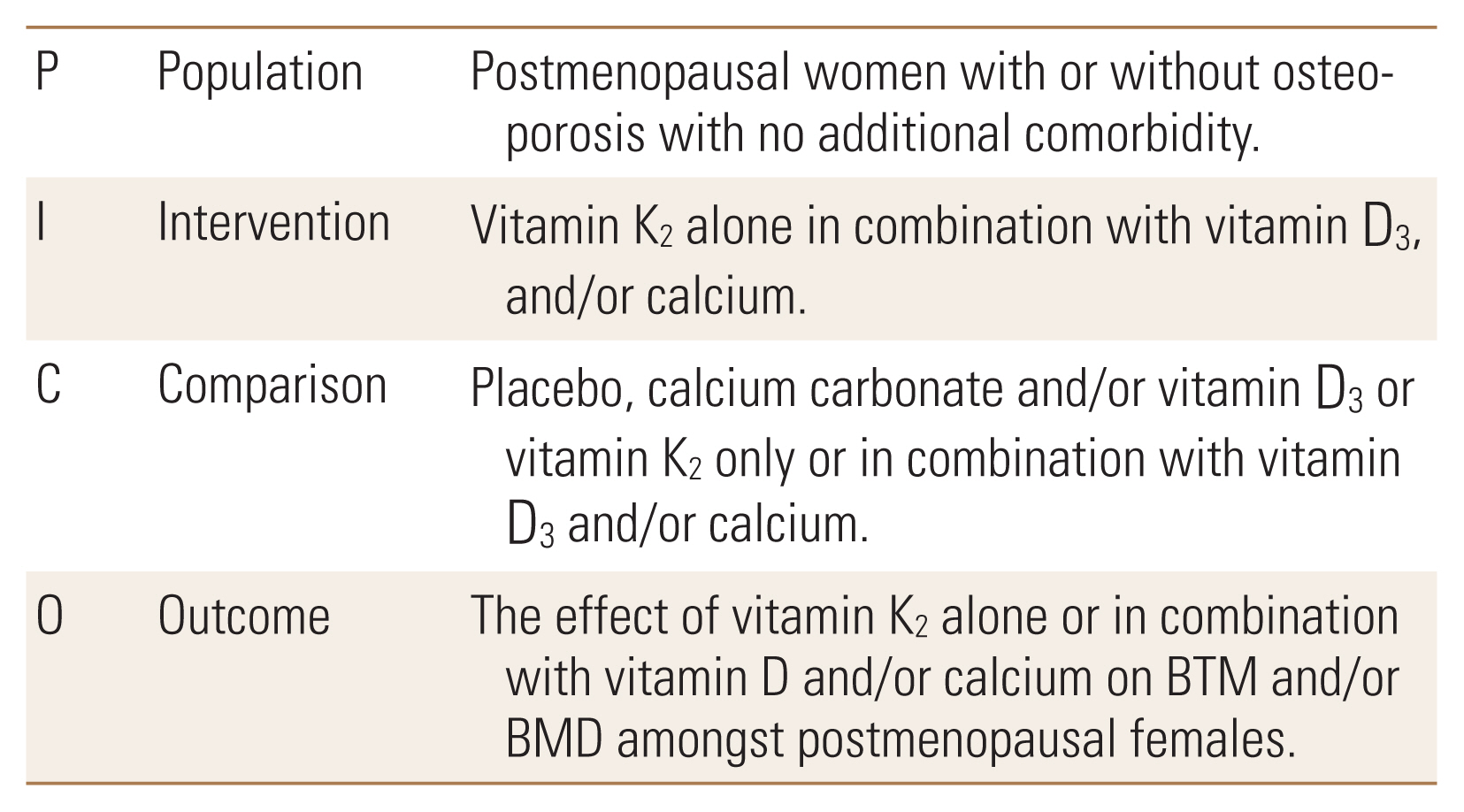
A systematic review of literature was performed based on the Preferred Reporting Items for Systematic Review (PRISMA) guidelines.[29] MEDLINE, ScienceDirect, PubMed, and Google Scholar, were searched using the key terms described, identified through PICO (Fig. 1), the review question, and initial scoping and MeSH search.

2. Study selection

Studies were included if they met the following inclusion criteria and were available as a full text:
  1. Population: postmenopausal women with/without osteoporosis with no additional comorbidity.

  2. Design: RCT

  3. Aim: evaluate the effect of vitamin K2 alone or in combination with vitamin D and/or calcium on BTM and/or BMD amongst postmenopausal females.

  4. Intervention: vitamin K2 alone or in combination with vitamin D3, and/or calcium.

  5. Comparator/s: placebo, calcium carbonate and/or vitamin D3 or vitamin K2 only or in combination with vitamin D3 and/or calcium.

  6. Publication language, and accessibility: published in English and can be sourced as a full text either directly from the journal by using the Higher Colleges of Technology Library and/or Lancaster University.

  7. Jadad score ≥2

3. Quality assessment

Jadad scale was used to assess quality of studies.[30,31] A score ≤2 indicates a low-quality design, while ≥3 indicates a high-quality design.[30,31] Three researchers calculated these scores, which were then validated by a fourth researcher.

4. Data extraction, and synthesis

A standardized document was used to extract data by 3 researchers, which was reviewed by a fourth researcher. Data were synthesized narratively, and results presented in a tabulation format.

RESULTS

The search process is displayed in Figure 2. Using key terms seen in Figure 2, 5,289 studies were identified from four databases. Titles were then screened for including key terms, and the inclusion criteria were applied by inspecting abstracts and/or full texts, resulting in including 9 studies.
All studies were RCT, and most of them were conducted in East Asia. The comparators across studies were not consistent, where some used placebo, while others used calcium carbonate and/or vitamin D3 or vitamin K2 only compared to a combination of vitamin K2 with vitamin D3. Different types and doses of vitamin K2 were used. Five out of the 9 studies used MK-4, 3 used 45 mg/day, and 1 used 1.5 mg/day. The remaining four studies used MK-7 in various doses ranging between 100 mcg and 45 mg/day. There are variations between the 2 types of vitamin K2, where MK-4 requires higher doses than MK-7 due to differences in the bioavailability.[32] Also, studies included had Jadad score between 2 and 4. Results are summarized in Table 1.

1. BMD

Table 2 provides a summary of the number of studies that investigated each parameter.
Purwosunu et al. [33] who conducted RCT on 63 participants, found that the treatment group (vitamin K2+calcium) had a significant positive change from baseline, unlike the comparator group (placebo+calcium), with a significant difference between both. Furthermore, Yasui et al. [34] who also conducted RCT on 30 participants, found that lumbar spine BMD reduced significantly in group 1 (vitamin K2), but insignificant in group 2 (vitamin K2+vitamin D3). However, the difference between groups was not tested.[34] Additionally, in Ushiroyama et al. [35] who conducted RCT, the vertebral BMD in groups 1 (vitamin K2) and group 2 (1-α hydro cholecalciferol) increased insignificantly, unlike the significant increase from baseline in group 3 (vitamin D3+ vitamin K2). However, group 4 (dietary therapy as control) showed a significant reduction from baseline. The difference between the groups was not tested.[35] Moreover, Jiang et al. [36] conducted a multi-center RCT on 213 participants, where the lumbar spine and trochanter BMD in groups 1 (vitamin K2+calcium) and 2 (Alfacalcidol+calcium) increased significantly from baseline, with no significant difference between groups. Also, there was no change in the femoral neck in both groups from baseline.[36]
Nevertheless, dual energy X-ray absorptiometry-BMD did not change in both groups in Knapen et al. [37] who has conducted RCT on 257 participants. Rønn et al. [38] has conducted RCT on 142 participants, where the tibia volumetric BMD (vBMD) did not change in group 1 (vitamin K2+calcium+vitamin D), yet percentages reduced significantly in group 2 (placebo+calcium+vitamin D), with a significant difference between groups. Furthermore, the percentages of radius vBMD in both groups were not different.[38] In addition, the changes in areal BMD were small and did not differ much between groups.[38]
On the other hand, Emaus et al. [39] who have conducted RCT, observed an insignificant reduction in BMD in total hip, femoral neck, lumbar spine, and total body in both groups (vitamin K2 and placebo) with a significant difference between groups.

2. OC variables

1) OC

Levels in group 1 in Ushiroyama et al. [35] went below baseline (P>0.05), while, in group 3, it increased significantly. Yet, the difference between groups was not tested.[35] In addition, levels in group 1 in Yasui et al. [34] reduced insignificantly, while in group 2 it dropped significantly from baseline, yet the difference between groups was not tested. Emaus et al. [39] also looked into the effect of vitamin K2 on N-mid OC, where the treatment group (vitamin K2) had more reduction from baseline than the placebo group, with a significant difference between both. Jiang et al. [36] found that the reduction was significant from baseline in both, but vitamin K2 group reduction was more profound. Also, the difference between the 2 groups was statistically significant.[36] Furthermore, group 1 in Rønn et al. [38] experienced a reduction in OC after 6 and 12 months, while in group 2 it remained almost static, with a significant difference between both. On the other hand, levels of groups 1 and 2 in Purwosunu et al. [33] increased from baseline, yet the difference from baseline was significant only in group 1, with no significant difference between both groups.

2) ucOC

In group 1 (vitamin K2+calcium) in Purwosunu et al. [33] ucOC reduced significantly from baseline, unlike in group 2 (placebo+calcium), with a significant difference between both groups. Koitaya et al. [40] who also conducted RCT for 40 participants, found that ucOC in group 1 (vitamin K2) reduced significantly, however, there was a significant increase in group 2 (placebo) from baseline. The difference between the 2 groups after 4 weeks was also statistically significant.[40] Also, Knapen et al. [37] found that ucOC in group 1 (vitamin K2+calcium+vitamin D) declined from baseline, while in group 2 (placebo+calcium+vitamin D) it increased, with a significant difference between the 2 groups. Emaus et al. [39] also showed a decline in both groups, but more profoundly and significantly in vitamin K2 receivers. Inaba et al. [41] who conducted RCT including 115 participants, found that in group 1 (vitamin K2) it reduced significantly from baseline, yet levels in group 2 (placebo) remained almost the same. Along the same line, ucOC in group 1 in Rønn et al. [38] reduced from baseline, while group 2 showed almost no changes, with a significant difference between both. Furthermore, both groups in Jiang et al. [36] had a significant reduction from baseline, but vitamin K2 receivers had a higher reduction.

3) Carboxylated OC (cOC)

cOC in group 1 (vitamin K2) in Knapen et al. [37] increased from baseline, unlike in group 2 (placebo), with a significant difference between both. Similar findings were seen in Emaus et al. [39] where cOC in both groups increased from baseline, but the increase was higher in group 1 (vitamin K2), with a statistically significant difference between both. Furthermore, in Inaba et al. [41] group 2 (placebo) showed an insignificant reduction from baseline, while there was an insignificant increase in group 1 (vitamin K2). Koitaya et al. [40] which explored γ-carboxylated OC (Gla-OC), found a significant increase from baseline in both groups, but more profoundly in group 1 (vitamin K2), with a significant difference between both groups only after 2 weeks.

4) ucOC:cOC ratio

In Inaba et al. [41] the ratio in group 1 (vitamin K2) increased significantly but remained almost the same in group 2 (placebo). However, ucOC/OC in group 1 in Rønn et al. [38] declined from baseline, compared to group 2, with a significant difference between both. Furthermore, in Jiang et al. [36] the levels reduced significantly in both groups, but the reduction was more profound in group 1.

3. γ-carboxyglutaminate Gla/creatinine (Cr)

This was only explored in Ushiroyama et al. [35] where levels in group 1 increased significantly, while, in group 3, the increase was insignificant. Yet, the difference between both groups was not tested.[35]

4. Total OC or Gla-OC/Gla-OC+ucOC ratio

The ratio in groups 1 and 2 in Koitaya et al. [40] increased significantly, but the increase was more profound in the vitamin K2 group. The difference between the groups after 4 weeks was significant.[40] Besides, findings from Knapen et al. [37] brought to light that the total OC in group 1 and group 2 remained almost the same after 12 months of use.

5. Bone-specific alkaline phosphatase or bone alkaline phosphatase (BAP)

BAP in group 1 (vitamin K2) in Yausi et al. [34] remained almost static, while it dropped significantly in group 2 (vitamin K2 and vitamin D3), yet the difference between both was not tested. Furthermore, in Emaus et al. [39] levels in group 1 (vitamin K2) did not change from baseline, while, in group 2, it declined, with no significant difference between both groups. In Knapen et al. [37] however, BAP in both groups increased in the same manner from baseline. Besides, in Rønn et al. [38] BAP in vitamin K2+calcium+ vitamin D receivers increased from baseline compared to placebo+calcium+vitamin D receivers, with a significant difference between both.

6. Procollagen type 1 N-terminal propeptide (PINP) and carboxyterminal propeptide of type I procollagen (PICP)

PINP in both groups in Rønn et al. [38] increased from baseline, while in Ushiroyama et al. [35] the levels fluctuated in vitamin K2 group, then stabilized below zero. The differences between results after 6, 12, and 18 months and baseline were significant.[35] However, the levels increased after 24 months among the combined therapy group (vitamin K2 and D3), where differences were significant after 18 and 24 months compared to baseline.[35]

7. C-terminal telopeptide of type I collagen (CTX), and crosslaps (CL), N-terminal telopeptide levels (NTX) OR NTX/Cr ratio, deoxypyridinoline (DPD), and urinary pyridinoline

CTX in both groups in Rønn et al. [38] increased slightly from baseline. Furthermore, CL reduced slightly from baseline in both groups in Emaus et al. [39]. Regarding NTX, levels increased from baseline in both groups in Knapen et al. [37] but more profoundly in the vitamin K2 group, with no significant difference between them. Koitaya et al. [40] however, found that the NTX/Cr ratio decreased insignificantly in both groups, with no significant difference between the groups. Regarding urinary pyridine, the levels increased significantly in groups 1 and 3 in Ushiroyama et al. [35] yet, the difference between the 2 was not tested. Yausi et al. [34] also explored the effect of vitamin K2 on urinary DPD, where levels in both groups reduced insignificantly, but the reduction was more profound in group 2, and the difference between both was not tested. Koitaya et al. [40] who explored DPD/Cr, found that levels in group 1 increased significantly, unlike group 2, with no significant difference between the 2 groups.

DISCUSSION

This is a review of 9 RCTs mainly conducted to explore the effect of vitamin K2 alone or in combination with other agents (vitamin D3 or calcium) on BTMs amongst postmenopausal females. Based on the studies, vitamin K2 has positive and negative effects on various bone parameters.

1. BMD

BMD increased from baseline in the groups that received vitamin K2.[33,35,36] However, the difference between groups was only significant in 1 study.[33] Furthermore, it was observed that in certain studies, the difference between groups was not tested,[35] yet a rationale was also not provided. Nevertheless, it could be due to the small sample sizes,[35] however, in some other studies, despite the small sample sizes, the difference was tested.[33] There should have been a discussion around the normal distribution and the appropriateness of the statistical tests in studies with small sizes.[33,35] Importantly, statistical significance does not translate into clinical significance,[42,43] as there might be changes from the baseline which are clinically but not statistically significant. In all 3 studies showing a significant increase from baseline, vitamin K2 was administered alongside another agent (calcium/vitamin D), which might have inflated the results.[33,35,36] A meta-analysis of 19 RCTs echoed this as a significant improvement in vertebral BMD amongst vitamin K2 receivers.[28] In some cases, the comparator was given an active ingredient rather than a placebo or alongside the placebo, which justifies the improvement in values.[36] On the other hand, some studies demonstrated the positive effect of vitamin K2 by stabilizing BMD and preventing deterioration.[37,38] As per literature, the lifetime risk of having at least 1 fracture reduced by 25% with the daily use of 800 IU vitamin D, 45 μgm vitamin K2, and 1,200 mg calcium, and that using vitamin K2 reduces the decline in BMD due to aging.[44] In fact, the low levels of vitamin K2 were associated with low BMD and increased risk of hip fracture.[45] On the other hand, in another 2 studies, the results reduced from the baseline, opposing the previous discussion.[34,39] Holistically, vitamin K2 is advantageous in either increasing BMD or preventing further deterioration in the majority of studies. And this goes in line with the results from a meta-analysis confirming the effectiveness of vitamin K2.[46]

2. OC

OC increases during postmenopausal osteoporosis and decreases post-therapy,[47-50] however in Purwosunu et al. [33], there was an increase from baseline in both groups, but significantly in the vitamin K2+calcium receivers, and this was not expected, since both groups received at least calcium.[33] However, since these were measures during postmenopause, this could have afflicted the findings.[47-50] Additionally, in Ushiroyama et al. [35], levels reduced amongst vitamin K2 receivers only, while there was an increase in the group that received a combination (vitamin K2 and vitamin D).[35] For group 1, this indicates an improvement in bone health yet results in groups are opposing what is expected, hence adding another layer of controversy.[35] Again, no rationale was provided by the study to justify these findings.[35] However, in another 4 studies, the positive effect of vitamin K2 was evident by reducing OC from baseline.[34,36,38,39] The positive effect of vitamin K2 on OC was also seen in literature, as vitamin K enhanced the accumulation of Gla OC, OC production, and mineralization induced by vitamin D3.[21,51] On the other hand, in Knapen et al. [37] OC in both groups remained static, although it was used for a long time (3 years). This can be viewed as a positive effect, though, as the prevention of deterioration during postmenopause means that it has the potential to be used prophylactically. When viewing all these findings holistically, it seems that vitamin K2 is useful for bone health, which also can be used prophylactically.

3. ucOC

An increase in ucOC indicates a deterioration in bone health through increasing bone resorption activity.[50,52] It has been disputed that a high ucOC level may be a marker of hip fracture risk in older women.[53,54] In all studies investigating ucOC, the groups that have used vitamin K2 had a reduction in ucOC, and in most of these studies, the reduction was significant, and the difference between the groups was also significant.[33,36-41] Despite the variations in doses, types of vitamin K2, intervention and the comparator groups, and duration of intervention, an overall reduction from baseline was observed, signposting a reduction in bone resorption activities.[33,36-41] Yet, interpretation should be made carefully, as in some studies where calcium and/or vitamin D were given to the comparator group, there was either a reduction or no changes from baseline.[33,36,38] Furthermore, in some studies, the intervention groups received vitamin K2 alongside other active ingredients, which might have inflated the findings.[33,36,38] Yet, in studies that used vitamin K2 alone against placebo, results were still positive.[37,39-41] This goes in line with the literature, where MK-4 recipients showed a reduction in ucOC within 2 weeks,[55] and ucOC was inversely associated with spinal BMD in healthy Korean women.[56]
An elevation cOC and Gla-OC reflects osteoblasts activity.[52] In 4 studies, vitamin K2 receivers’ levels increased,[37, 39-41] meaning an increase in osteoblasts’ activity.[52] Despite the small sample size and a smaller dose of MK-7, vitamin K2 was still effective but not significantly.[41] Yet, as mentioned earlier, being statistically significant does not mean being clinically significant. Moreover, in Ushiroyama et al. [35] vitamin K2 receivers showed a significant increase in γ-carboxyglutaminate Gla/Cr, unlike vitamin K2+D receivers, which had a slight and insignificant increase from baseline. Unfortunately, it was not possible to know why, as it should demonstrate more improvement due to the dual therapy.[35] These overall positive findings go well with the literature, where the administration of 45 mg MK-4 increased OC and reduced fracture, suggesting that the co-administration of vitamin K with bisphosphonates might improve the osseous effect of bisphosphonate.[57-59] However, only in Koitaya et al. [40] the placebo group experienced an increase in the levels, and it was not possible to understand why. Overall, vitamin K2 seems to be effective in elevating osteoblast activity. Hence, it would be a good supplement taken by postmenopausal women.

4. ucOC:cOC or cOC:ucOC ratio

The ratio increases when vitamin K levels are low.[60,61] In 2 studies, the groups receiving vitamin K2 alongside calcium only and/or with vitamin D experienced a more profound reduction.[36,38] This goes in line with the literature, where the ratio improved with the use of MK-7.[62] However, this was not the case in Inaba et al. [41] as levels increased among vitamin K2 receivers, which might be due to the low dose of MK-7 (100 mcg). This is a clear indication that the pharmacokinetic profile of vitamin K2 needs to be further investigated. Additionally, in Inaba et al. [41] vitamin K2 was used alone, unlike in the other 2 studies, indicating that the ratio might be sensitive to other agents. Koitaya et al. [40] which explored Gla-OC/Gla-OC+ucOC ratio, found that levels increased from baseline more profoundly among vitamin K2 receivers compared to the placebo, despite the small dose and short duration of the intervention. Overall, the ratio seems to be sensitive to vitamin K’s presence in blood, and this can be used to monitor patients or for future research.

5. BAP

BAP increases in patients with bone diseases.[63] In 2 studies, the groups that have used vitamin K2 only showed no difference from baseline,[34,39] which means no deterioration in bone health. These findings were not supported by literature, where BAP decreased among glucocorticoid users, but not in the group that used vitamin K2 with glucocorticoids.[64] Findings from Knapen et al. [37] echoed the outcome from literature, where levels increased with the administration of vitamin K2, and this could be reflecting bone health status during postmenopause. Overall, whenever a combination was used (vitamin K2+vitamin D or calcium+vitamin D), the results reduced significantly, with an exception to the group using triple therapy (vitamin K2+vitamin D+calcium), which showed an increase from baseline. Yet, a rationale was not provided.[34,37-39] In general, findings are not conclusive regarding BAP, however, in 2 studies, the effectiveness was demonstrated by preventing deterioration, which means it could be useful prophylactically.

6. PINP, PICP, and CTX

PINP is derived from collagen type I, the most abundant form of collagen found in bone and synthesized by osteoblasts.[65,66] Since these are generated from newly synthesized collagen, they are considered quantitative measures of newly formed type I collagen.[65,66] PICP is a specific marker of proliferating osteoblasts and fibroblasts, therefore, it is a marker of bone formation.[48] Furthermore, elevated levels of CTX indicate an increase in bone resorption.[67] PINP, PICP, and CTX in the groups that used a combination (vitamin D and calcium or vitamin K2, vitamin D, and calcium) increased from baseline.[38] This increase in PINP and CTX reflects osteoblast and osteoclast activity, respectively, indicating an increase in bone remodeling activity. PICP remained the same in Ushiroyama et al. [35] among vitamin K2 consumers, while it increased significantly among vitamin K2+D users, which might be due to the additive effect of the combination. CL in Emaus et al. [39] were reduced mildly and insignificantly in both groups (vitamin K2 and placebo). The reduction in both groups was equivalent, and it was not possible to rationalise the lack of information provided on the emergence of such results.[39] As these parameters were not explored by plenty of the included studies, the results do not seem conclusive.

7. NTX or NTX/Cr ratio

An elevation in the NTX or NTX/Cr ratio levels indicates unbalanced remodeling.[65,66,68] In Knapen et al. [37] levels in both groups increased, with a higher increase seen among vitamin K2 users. In the vitamin K2 group, this could indicate an increase in osteoblast activity; however, this means unbalanced remodeling in the placebo group. In Koitaya et al. [40] there was an insignificant reduction from baseline, indicating a reduction in the imbalance or achieving equilibrium, a positive outcome. However, as these were tested after 4 weeks, results might not be accurate.[40,68,69]

8. DPD

DPD is high in patients who are having bone disorders such as osteoporosis.[70] In 2 studies, the levels reduced from baseline, and this reduction was significant in one study,[35] and insignificant in the other.[34] Surprisingly, groups that used a combination (vitamin K2+D) showed conflicting results, where values increased significantly from the baseline in one study, which might be due to the short duration (4 weeks),[35] while it reduced insignificantly in another study.[34] In Koitaya et al. [40] the levels increased from baseline among vitamin K users. The results might also have been afflicted by participants’ age and the expected change in bone status. Furthermore, it seems that the short duration does not allow a proper assessment of DPD, since it requires time (more than 4 weeks) to restore or treat bone health.[40,68,69]

9. Impact of duration, sample sizes, populations, and choices of comparator groups

There were variations between studies in terms of the duration of interventions (2 weeks-3 years), which might affected the results. For instance, NTX or NTX/Cr was tested after 4 weeks, but it needs at least 6 months for accurate assessment.[40,68,69] DPD also seems to be affected by the duration of the intervention and discussed earlier results.[40,68,69] The variations were also seen in vitamin K2 doses and the comparator groups, ranging between placebo, vitamin D and/or calcium. For example, when the comparator is given vitamin K2 with vitamin D, findings might be inflated,[34] and as the comparator groups were diverse, drawing comparisons between studies was not an easy exercise. Not all findings were significant, which might be due to the sample sizes, given the relationship between sample size, confidence intervals, and P values. Hence, an accurate interpretation of results will require effect sizes to be reported.[71,72] Most studies were conducted in East Asian countries, meaning that generalization should be exercised with extreme caution given their small body frame.

10. Situations afflicting BTMs

The OC, the ucOC and cOC, have extra-skeletal roles, such as their roles in glucose metabolism, hence levels might be altered among diabetics, which is common at the age of menopause.[73,74] Furthermore, a decrease in Glu-OC might be a symptom of insulin resistance and the appearance of markers of low-grade inflammation accompanying obesity.[75] Moreover, ucOC/cOC ratio is altered in hemodialysis, resulting in losing its significance.[76] Yet, studies included in this systematic review have not incorporated patients with conditions that might alter BTMs. Additionally, certain BTMs such as PINP might be seen in other locations such as the skin, dentin, cornea, vessels, fibrocartilage, and tendons.[77] However, most of the non-skeletal contribute very little to the circulating propeptide pool.[77]

11. Vitamin K2 use challenges

The administration of vitamin K2 is not without obstacles, as some factors interfering with its absorption, such as: antibiotic use, Dilantin, low-fat diet and fat blocking supplements, bile acid sequestrants, orlistat, Xenical, and olestra, mineral oil, and preservative butylated hydroxytoluene, gastrointestinal tract diseases, liver diseases, and estrogen drugs.[78] Also, green leaves are a potential source of interaction.[78,79] Furthermore, both types o vitamin K interact with warfarin and affects the International Normalized Ratio.[78,79]

12. Limitations

One of the limitations of this review is the small number of studies indicating that evidence is scarce. Concerning the included studies, several limitations were encountered, such as the variation in doses, types of vitamin K2 used, duration of intervention, and the product given to the groups. Furthermore, the small sizes of populations might have affected the statistical comparison between groups. Additionally, the findings’ generalizability remains questionable, as most studies (except 2) were conducted in either Japan or Indonesia (East Asia), which are known to have smaller body frames. Also, not all studies are of high quality, however, it was not possible to exclude given the limited number of suitable studies.

CONCLUSION

Vitamin K2 might be beneficial as an added therapy in managing and preventing postmenopausal osteoporosis, as demonstrated on BMD, OC, ucOC, cOC, and Gla-OC levels. Yet, findings were not conclusive when testing PINP, PICP, CTX, BAP, DPD, ucOC:cOC or cOC:ucOC ratio, NTX or NTX/Cr ratio. The administration of vitamin K2 alongside vitamin D and calcium rather than each 1 alone is presumed to be advantageous as per most studies included in this study. Yet, this is not to say that vitamin K2 is to replace existing therapy since taking vitamin K2 routinely is not internationally recommended for postmenopausal women with osteoporosis. In addition, a proper understanding of the pharmacokinetics and pharmacodynamics of vitamin K2 is a cornerstone. Consequently, more studies are needed using standardized doses and types of vitamin K2 to ameliorate controversy related to the pharmacokinetics and pharmacodynamics profiles. Future studies should also include a wider range of BTMs to enable reaching consensus. Besides, as elucidated earlier, the use of vitamin K2 should be carried out with caution, especially when certain medications are on board or when the patient has comorbidities.

Supplementary Information

Supplementary Appendix 1
Bone markers
OC variables including:
  • - OC

  • - ucOC

  • - cOC or plasma Gla-OC

  • - ucOC: cOC or cOC: ucOC ratio

  • - Total OC or Gla-OC/Gla-OC+ucOC ratio

  • - Gla/Cr

  • - OC is a highly sensitive marker for bone formation, it is a tissue specific marker, lacks interpersonal variations, and reflects the osteoblastic activity.[43-46] OC is known to increase during postmenopausal osteoporosis and decreases post-therapy.[43-46] The standard immunochemical assays for evaluating osteoblastic activity are intact OC (amino acids 1-49) and N-mid OC (amino acids 1-43).[43-46] N-mid OC is apparently more stable when compared to intact OC due to protease cleavage between amino acids 43 and 44.[43-47]

  • - cOC gets elevation in the levels of carboxylated variants of OC is an indicator of bone formation activities (osteoblast).[43-47]

  • - ucOC is produced through an imperfect γ-carboxylation, which is a bone marker reflecting the bone resorption activities as well as the vitamin K status in the bone.[46,48] In vitamin K deficiency, as the levels of c-carboxylation reduces, this does not enable a large portion of OC to undergo the complete carboxylation process, hence it is being termed as the ucOC.[49] A negative association between serum levels of ucOC and BMD at the hip has been reported,[50] and it has been disputed that a high ucOC level may be a marker of hip fracture risk in elderly women.[50,51]

  • - γ-carboxyglutaminate protein is involved in the local control of calcium deposition in mineralized tissue, and is often high in patients with osteoporosis.[22,46,48]

  • - The ucOC: cOC ratio as well can be used as an indicator for the status of vitamin K, where an elevation in the ratio indicates low level of vitamin K.[52,53]

  • - γ-carboxyglutaminate Gla/Cr protein is involved in the local control of calcium deposition in mineralized tissue, and is often high in patients with osteoporosis. [22,46,48]

Bone-derived alkaline phosphatase:
  • - BAP or used interchangeably with serum BAP or BSAP

  • - These are bone specific isoforms of alkaline phosphate reflecting the biosynthetic activity of bone-forming cells, which is found to be high in diseases or conditions such as Paget, osteomalacia and osteoporosis.[54]

Collagen peptides:
  • - PINP

  • - PICP

  • - PINP are derived from collage type I, the most abundant form of collagen found in bone.[55,56] In bone, collagen is synthesised by osteoblasts in the form of pre-procollagen which are characterized by having the PINP and PICP.[55,56] Since these are generated from newly synthesised collagen, they are considered quantitative measures of newly formed type I collagen.[55,56]

  • - PICP is a specific marker of proliferating osteoblasts and fibroblasts, therefore is a marker of bone formation.[44]

Collagen degradation products:
  • - Cross-linked NTX or aminoterminal cross-linked telopeptide of type I collagen

  • - Cross-linked CTX or C-terminal cross-linked telopeptide of type I collagen or CL

  • - DPD

  • - Urinary pyridinoline

  • - NTX molecules are mobilized from bone by osteoclasts and subsequently excreted in the urine, and an increase in the levels of NTX indicates an increase in the bone turnover, more specifically it is an indication of unbalanced remodeling (osteoblasts and osteoclasts) which is usually seen in osteoporosis.[55-57] NTX is also expressed as a ratio of Cr.

  • - During the bone resorption, osteoclasts secrete a mixture of acid and neutral proteases that degrade the collagen fibrils into molecular fragments CTX.[58] Elevated levels of CTX indicate increased bone resorption, and increased levels are associated with osteoporosis, osteopenia, Paget disease, hyperthyroidism, and hyperparathyroidism.[58]

  • - CL, which measures the degradation of CTX of type I collagen.

  • - The bone turnover process can be assessed by exploring the degradation of bone collagen such as the pyridinoline and DPD, which are formed during the maturation of bones.[59,60] During bone resorption, when mature bone collagen is degraded, these compounds get released and then eliminated via the kidneys.[59,60] The levels are found to be higher in patients with osteoporosis in comparison to healthy individuals.[61]

OC, osteocalcin; ucOC, undercarboxylated OC; cOC, carboxylated OC; Gla-OC, γ-carboxylated OC; Cr, creatinine; BAP, bone alkaline phosphatase; BSAP, bone-specific alkaline phosphatase; PINP, procollagen type 1 N-terminal propeptide; PICP, propeptide of type I procollagen; NTX, N-terminal telopeptide levels; CTX, C-terminal telopeptide of type I collagen; CL, crosslaps; DPD, deoxypyridinoline; N-mid, N-terminal/midregion.

DECLARATIONS

Funding

This research received no specific grant from any funding agency in the public, commercial, or not-for-profit sectors.

Ethics approval and consent to participate

Not applicable.

Conflict of interest

No potential conflict of interest relevant to this article was reported.

Fig. 1
Population, Intervention, Comparison, Outcome (PICO). BTM, bone turnover marker; BMD, bone mineral density. [Modified from “PICO, PICOS and SPIDER: a comparison study of specificity and sensitivity in three search tools for qualitative systematic reviews.”, by Methley AM, et al., 2014, BMC Health Serv Res, 14, p. 579. Copyright 2014 by the BioMed Central. Modified with permission].
jbm-2021-28-1-11f1.jpg
Fig. 2
Preferred Reporting Items for Systematic Reviews and Meta-Analyses flow chart.
jbm-2021-28-1-11f2.jpg
Table 1
Results gleaned from randomized controlled trials
References Year Jadad scale Groups and sample size Design Duration of intervention Efficacy parameters or measures Main outcomes in the efficacy parameter
Knapen et al. [37] 2007 2 Total number (N=257): group 1 (vitamin K2 once daily): N=133, Group 2 (placebo): N=124 Randomized control study 3 years BMD (this was measured after 1, 2, and 3 years of starting the treatment/placebo), BMC, tOC, ucOC, cOC, BAP, sNTX, free DPD. These were measured at baseline, and then in months 3, 6, 12 and 36. However, all results were reported after 12 months of use. BMD (g/cm2): no effect at any of the sites measured; BMC (g): group 1 and 2 levels in group 1 decreased at a significantly lower rate than in the placebo one (P<0.05). OC variables: (1) cOC (ng/mL): group 1 increased by about 0.5 ng/mL, group 2 reduced by about 2 ng/mL, difference between groups: significant; (2) ucOC (ng/mL): group 1 declined by about>2 ng/mL, group 2 increased by about 1 ng/mL, difference between groups: significant; (3) tOC (ng/ mL): the levels in both groups remained almost the same (14 ng/mL). BAP reported in U/l: the levels in groups 1 and 2 increased by around 4 U/l. Collagen degradation products: (1) NTX (nM): group 1 increased by about 2 nM, group 2 increased by about 1 nM, difference between groups: insignificant; (2) DPD/creat (nmol/mmol): levels in both groups reduced by approximately 2 nmol/mmol.
Emaus et al. [39] 2010 5 Total number (N=299): group 1 (vitamin K2 once daily): N=153, group 2 (placebo): N=146 Randomized, double-blind, placebo-controlled trial 12 months BMD, sOC, CL, ucOC, cOC, BAP. These were measured at baseline and after 12 months. BMD (g/cm2): (1) Total hip: group 1 reduced by about 0.004 g/cm2, group 2 reduced by about 0.003 g/cm2, difference between groups: insignificant; (2) Femoral neck: group 1 reduced by approximately 0.004 g/cm2, group 2 reduced by 0.005 g/cm2, difference between groups: insignificant; (3) Lumbar spine: group 1 reduced by 0.006 g/cm2, group 2 declined by 0.006 g/cm2, difference between groups: insignificant; (4) Total body: group 1 reduced by 0.010 g/cm2, group 2 reduced by 0.009 g/cm2, difference between groups: insignificant. OC variables: (1) N-mid OC (ng/mL): group 1 reduced by about 4.78 ng/mL, group 2 reduced by 1.66 ng/mL, difference between groups: significant; (2) cOC (ng/mL): group 1 increased by about 5.56 ng/mL, group 2 it increased by around 1.70 ng/mL, difference between groups: significant; (3) ucOC (ng/mL): group 1 reduced by 1.92 ng/mL, group 2: reduced by 0.24 ng/mL, difference between groups: significant. BAP reported in μg/L: group 1 no change, group 2 reduced by 1.11 μg/L, difference between groups: insignificant. Collagen degradation products (CL reported in ng/mL): the levels in group 1 and 2 declined by about 0.06 after 12 months of use.
Inaba et al. [41] 2015 5 Total number (N=115): group 1 (vitamin K): N=58, group 2 (placebo): N=57 Double-blinded randomized controlled trial 84 days cOC, ucOC, cOC/ucOC ratio. These were measured at baseline, and after 28, 56, 84, and 112 days. Importantly, the vitamin K2/placebo were only ingested for a period of 84 days. OC variables: (1) cOC (ng/mL): group 1 increased from baseline by about 2.29 ng/mL, group 2 reduced by about 2 ng/mL, difference between groups: insignificant; (2) ucOC (ng/mL): group 1 reduced significantly by 1.5 ng/mL, group 2 no change; (3) cOC/ucOC ratio: group 1 increased significantly by about 0.5, group 2 it remained almost the same.
Rønn et al. [38] 2016 3 Total number (N=142): group 1 (vitamin K, calcium, and vitamin D): N=71, group 2 (placebo, calcium, and vitamin D): N=71 Randomized, placebo-controlled, double-blinded clinical trial 1 year BMD, OC, ucOC, PINP, BAP, CTX. These were measured at baseline, and after 3, 6, 9, and 12 months. vBMD of the tibia and radius (in percentage): (1) Tibia: group 1 no change, group 2 decreased significantly by 3.5±8.6%, difference between groups: significant; (2) Radius: no changes in both groups. aBMD (in percentages): changes after 12 months were small and did not differ between the groups. OC variables: (1) OC (changes in the mean represented in percentages): group 1 reduced by 25% after 6 and 12 months, group 2 remained almost static, difference between groups: significant; (2) ucOC (changes in the mean represented in percentages): group 1 reduced from baseline by about 80%, group 2 almost no changes, difference between groups: significant; (3) ucOC/OC ratio (changes in the mean represented in percentages): group 1 reduced by about 55%, group 2 almost no changes, difference between groups: significant. BAP (changes in the mean represented in percentages): group 1 increased by about 5%, group 2: slight reduction, difference between groups: significant. Collagen peptides (PINP, changes in the mean represented in percentages): both groups showed an increase in the levels by about 5% from baseline. Collagen degradation products (CTX, changes in the mean represented in percentages): both groups showed a slight elevation in the levels of CTX by about 5% after 12 months.
Jiang et al. [36] 2014 3 Total number (N=213): group 1 (vitamin K2 and calcium once daily): N=108, group 2 (placebo and calcium carbonate once daily): N=105 Multi-center, randomized, double-blinded, double-dummy, positive drug controlled study 1 year BMD, OC, ucOC, ucOC/OC ratio. These were measured at baseline, and then in months 6 and 12. BMD (in percentage): (1) Lumbar spine: group 1 increased significantly by 1.2% after 12 months of use, group 2 increased significantly 2.2% after 12 months of use, difference between groups: insignificant; (2) Trochanter: group 1 increased significantly by 2.7% after 12 months of use, group 2 increased significantly by 1.8% after 12 months of use, difference between groups: insignificant; (3) Femoral neck: no significant changes in both groups from baseline. OC variables: (1) OC and ucOC (in percentage): group 1 OC and ucOC decreased significantly by 38.7% and 82.3%, respectively after 12 months, group 2 OC and ucOC decreased significantly by 25.8% and 34.8%, respectively after 12 months, difference between groups: significant; (2) ucOC/OC ratio (in percentage): the ratio in both groups reduced significantly after treatment, but the reduction was more profound in group 1.
Purwosunu et al. [33] 2006 4 Total number (N=63): group 1 (vitamin K2 and calcium carbonate once daily): N=30, group 2 (placebo and calcium carbonate once daily): N=33 Randomized double-blinded control study 48 weeks BMD, OC, ucOC. These were measured at baseline, and then in week 24 and 48. BMD of lumbar bone (in percentage): group 1 changed positively by 1.74±0.43%, group 2 changed negatively by −0.18±0.24%, difference between groups: significant. OC variables: (1) OC (ng/mL reported in mean±SD): group 1 increased significantly from 15 ng/mL to 23.1±13.6 ng/mL, group 2 increased insignificantly from about 20 ng/mL to 21.5±12.4 ng/mL, difference between groups: significant; (2) ucOC (ng/mL reported in mean±SD): group 1 reduced significantly from a little above 6 ng/mL to 2.6±2.15 ng/mL, group 2 reduced insignificantly from a little less than 6 ng/mL to 4.9±0.8 ng/mL, difference between groups: significant.
Yasui et al. [34] 2006 2 Total number (N=30): group 1 (vitamin K2 45 mg once daily): N=16, group 2 (vitamin K2 45 mg and vitamin D3 0.75 μg once daily): N=14 Randomized control study 2 years BMD at the lumbar and spine, OC, ucOC, DPD, BAP. These parameters were measured before the intervention, and then after the 1st year and 2nd year of intervention. The difference between groups in all these parameters was not tested. BMD (g/cm2): (1) Lumbar spine: group 1 reduced significantly from 0.743+0.052 to 0.685+0.038 g/cm2 after 2 years, group 2 declined insignificantly from 0.728+0.056 to 0.707+0.058 g/cm2 after 2 years. OC variables: (1) OC (ng/ mL): group 1 reduced insignificantly from 4.2+2.3 to 3.8+3.4 ng/ mL after 2 years, group 2 reduced significantly from 6.3+4.1 to 2.7+1.9 ng/mL after 2 years. BAP reported in U/L: group 1 remained almost the same, group 2 reduced significantly from 32.6+10.2 to 29.4+8.2 U/L after 2 years. Collagen degradation products (DPD reported in nM/mM Cr): group 1 reduced insignificantly from 9.4+2.6 to 8.0+3.3 nM/mM Cr after 2 years, group 2 reduced insignificantly from 11.7+6.5 to 5.8+0.7 nM/mM Cr after 2 years.
Ushiroyama et al. [35] 2002 2 Total number (N=126): group 1 (vitamin K2 45 mg): N=30, group 2 (vitamin D3, 1 − α hydroxycholecalciferol 1 μg): N=32, group 3: (combination): N=31, Group 4 (controls): N=33 Randomized control study 2 years BMD at the lumber spine, PICP, OC, Gla/Cr, urinary pyridinoline, blood coagulation profile “APTT, AT-III, fibrinogen and plasminogen”. These parameters were measured before the intervention, and after 6, 12, 24, and 48 months. The difference between groups in all these parameters was not tested. The BMD was compared between all four groups, while OC, Gla/Cr, urinary pyridinoline and PICP was compared between group 1 and 3. BMD g/cm2 reported as mean±SD: group 1 increased insignificantly by 0.012 g/cm2 after 24 months, group 2 increased insignificantly by 0.05 g/cm2 after 24 months, group 3 increased significantly by 0.025 g/cm2 after 24 months, group 4 reduced significantly by 0.035 g/cm2 after 24 months. OC variables: (1) Gla/Cr (nmol/MCM. Cr.) reported in percentages: group 1 increased significantly by 56.8% after 24 months, group 3 increased insignificantly by almost 10% after 24 months; (2) Intact OC (ng/mL) reported in percentages: group 1 initially increased, then went below baseline (P>0.05) after 24 months, group 3 increased significantly by 10% (P<0.05) after 24 months. Collagen peptides (PICP reported in percentages): group 1 initially increased, then decreased significantly (P<0.05) after 24 months, group 3 increased significantly by 24.2% (P<0.05) after 24 months. Collagen degradation products (urinary pyridinoline reported in percentages): group 1 increased significantly by 50% after 24 months, group 3 increased significantly by 84.5% after 24 months.
Koitaya et al. [40] 2009 4 Total number (N=40): group 1 (vitamin K2 1.5 mg once daily): N=20, group 2 (placebo): N=20 Randomized double-blinded placebo control trial 4 weeks Serum concentration of PK and MK-4, BAP, ucOC, Gla-OC, ratio of Gla-OC/Gla-OC+ucOC, urine free DPD, serum OH(25)D3. These parameters were measures before the intervention, and then after 2 and 4 weeks. OC variables: (1) ucOC (ng/mL) reported in digits: group 1 reduced significantly by 1.5 ng/mL, group 2 increased significantly by 0.4 ng/mL, difference between groups: significant only after 4 weeks; (2) Gla-OC (ng/mL): group 1 and 2 increased significantly from baseline (P<0.05), yet, the increase was more profound in vitamin K2 receivers, difference between groups: significant only after 4 weeks; (3) Gla-OC/Gla-OC+ucOC ratio: group 1 increased significantly by 0.1 after 4 weeks, group 2 remained almost the same after 4 weeks, difference between groups: significant only after 4 weeks. BAP reported in U/L: group 1 reduced significantly by 3.9 U/L, group 2 reduced significantly by 3.5 U/L. Collagen degradation products: (1) DPD/creatitine (nmol/mmoL Cr): group 1 increased significantly by 0.4 nmol/mmoL Cr, group 2 it increased insignificantly by 0.9 nmol/mmoL Cr, difference between groups: insignificant (2) NTX/Cr (nmol BCE/mmol Cr): group 1 increased significantly by 1.6 nmol/mmol Cr, group 2 decreased insignificantly by 1.1 nmol/mmoL Cr, difference between groups: insignificant.

BMD, bone mineral density; aBMD, areal bone mineral density; vBMD, volumetric bone mineral density; BMC, bone mineral content; OC, osteocalcin; sOC, serum osteocalcin; tOC, total osteocalcin; cOC, carboxylated osteocalcin; ucOC, undercarboxylated osteocalcin; BAP, bone-specific alkaline phosphate; NTX, N-terminal telopeptide levels; DPD, deoxypyridinoline; CL, crosslaps; SD, standard deviation; PINP, procollagen type 1 N-terminal propeptide; PICP, carboxyterminal propeptide of type I procollagen; CTX, C-terminal telopeptide of type I collagen; Gla, γ-carboxyglutaminate; APTT, activated partial thromboplastin time; AT-III, antithrombin III; PK, phylloquinone; MK-4, menaquinone-4; DPD, deoxypyridinoline; OH(25)D3, 25-hydroxy-vitamin D3; BCE, bone collagen equivalents; Cr, creatinine.

Table 2
Number of studies exploring each parameter
Parameter Number of studies References
BMD 7 Yasui et al. [34], Ushiroyama et al. [35], Jiang et al. [36], Knapen et al. [37], Rønn et al. [38], Emaus et al. [39], Koitaya et al. [40]
OC 9 Yasui et al. [34], Ushiroyama et al. [35], Jiang et al. [36], Knapen et al. [37], Rønn et al. [38], Emaus et al. [39], Koitaya et al. [40], Inaba et al. [41], Kazdin [42]
Gla/Cr and Gla-OC/Gla-OC+ucOC ratio 2 Ushiroyama et al. [35], Jiang et al. [36]
ucOC: cOC or cOC: ucOC ratio 3 Emaus et al. [39], Koitaya et al. [40], Kazdin [42]
BAP and BSAP 4 Ushiroyama et al. [35], Knapen et al. [37], Rønn et al. [38], Emaus et al. [39]
PINP and PICP 3 Jiang et al. [36], Rønn et al. [38], Emaus et al. [39]
CTX and CL 2 Rønn et al. [38], Emaus et al. [39]
NTX (nM) or NTX/Cr ratio 2 Knapen et al. [37], Inaba et al. [41]
DPD and urinary pyridinoline 3 Ushiroyama et al. [35], Jiang et al. [36], Inaba et al. [41]

BMD, bone mineral density; OC, osteocalcin; Gla, γ-carboxyglutamate; ucOC, undercarboxylated osteocalcin; BAP, bone alkaline phosphate; PINP, procollagen type 1 N-terminal propeptide; PICP, carboxyterminal propeptide of type I procollagen; CTX, C-terminal telopeptide of type I collagen; CL, crosslaps; NTX, N-terminal telopeptide levels; DPD, deoxypyridinoline.

REFERENCES

1. Wells BG, DiPiro JT, Schwinghammer TL, et al. Pharmacotherapy handbook. 10th ed. New York, NY: McGraw-Hill Education; 2017.

2. Delmas PD, Fraser M. Strong bones in later life: luxury or necessity? Bull World Health Organ 1999;77:416-22.
pmid pmc
3. Kanis JA, Melton LJ 3rd, Christiansen C, et al. The diagnosis of osteoporosis. J Bone Miner Res 1994;9:1137-41. http://dx.doi.org/10.1002/jbmr.5650090802 .
crossref pmid
4. National Osteoporosis Foundation. 2017 Annual report 2017 [cited by 2018 Mar 24]. Available from: https://www.nof.org/ .

5. Gullberg B, Johnell O, Kanis JA. World-wide projections for hip fracture. Osteoporos Int 1997;7:407-13. http://dx.doi.org/10.1007/pl00004148 .
crossref pmid
6. Sambrook P, Cooper C. Osteoporosis. Lancet 2006;367:2010-8. http://dx.doi.org/10.1016/s0140-6736(06)68891-0 .
crossref pmid
7. Zeind CS, Carvalho MG. Applied therapeutics. 11th ed. Philadelphia, PA: Wolters Kluwer Health; 2017.

8. Leibson CL, Tosteson AN, Gabriel SE, et al. Mortality, disability, and nursing home use for persons with and without hip fracture: a population-based study. J Am Geriatr Soc 2002;50:1644-50. http://dx.doi.org/10.1046/j.1532-5415.2002.50455.x .
crossref pmid
9. Garnero P, Sornay-Rendu E, Chapuy MC, et al. Increased bone turnover in late postmenopausal women is a major determinant of osteoporosis. J Bone Miner Res 1996;11:337-49. http://dx.doi.org/10.1002/jbmr.5650110307 .
crossref pmid
10. Thorne Research, Inc. Vitamin K2. Monograph. Altern Med Rev 2009;14:284-93.
pmid
11. Plaza SM, Lamson DW. Vitamin K2 in bone metabolism and osteoporosis. Altern Med Rev 2005;10:24-35.
pmid
12. Iwamoto J, Sato Y, Takeda T, et al. High-dose vitamin K supplementation reduces fracture incidence in postmenopausal women: a review of the literature. Nutr Res 2009;29:221-8. http://dx.doi.org/10.1016/j.nutres.2009.03.012 .
crossref pmid
13. Beulens JW, Booth SL, van den Heuvel EG, et al. The role of menaquinones (vitamin K2) in human health. Br J Nutr 2013;110:1357-68. http://dx.doi.org/10.1017/s0007114513001013 .
crossref pmid
14. Shearer MJ, Fu X, Booth SL. Vitamin K nutrition, metabolism, and requirements: current concepts and future research. Adv Nutr 2012;3:182-95. http://dx.doi.org/10.3945/an.111.001800 .
crossref pmid pmc
15. Ferland G. The vitamin K-dependent proteins: an update. Nutr Rev 1998;56:223-30. http://dx.doi.org/10.1111/j.1753-4887.1998.tb01753.x .
crossref pmid
16. Hauschka PV, Lian JB, Cole DE, et al. Osteocalcin and matrix Gla protein: vitamin K-dependent proteins in bone. Physiol Rev 1989;69:990-1047. http://dx.doi.org/10.1152/physrev.1989.69.3.990 .
crossref pmid
17. Lombardi G, Perego S, Luzi L, et al. A four-season molecule: osteocalcin. Updates in its physiological roles. Endocrine 2015;48:394-404. http://dx.doi.org/10.1007/s12020-014-0401-0 .
crossref pmid
18. Booth SL, Centi A, Smith SR, et al. The role of osteocalcin in human glucose metabolism: marker or mediator? Nat Rev Endocrinol 2013;9:43-55. http://dx.doi.org/10.1038/nrendo.2012.201 .
crossref pmid
19. Urayama S, Kawakami A, Nakashima T, et al. Effect of vitamin K2 on osteoblast apoptosis: vitamin K2 inhibits apoptotic cell death of human osteoblasts induced by Fas, proteasome inhibitor, etoposide, and staurosporine. J Lab Clin Med 2000;136:181-93. http://dx.doi.org/10.1067/mlc.2000.108754 .
crossref pmid
20. Gundberg CM, Lian JB, Gallop PM, et al. Urinary gamma-carboxyglutamic acid and serum osteocalcin as bone markers: studies in osteoporosis and Paget’s disease. J Clin Endocrinol Metab 1983;57:1221-5. http://dx.doi.org/10.1210/jcem-57-6-1221 .
pmid
21. Koshihara Y, Hoshi K. Vitamin K2 enhances osteocalcin accumulation in the extracellular matrix of human osteoblasts in vitro. J Bone Miner Res 1997;12:431-8. http://dx.doi.org/10.1359/jbmr.1997.12.3.431 .
crossref pmid
22. Kim M, Na W, Sohn C. Vitamin K1 (phylloquinone) and K2 (menaquinone-4) supplementation improves bone formation in a high-fat diet-induced obese mice. J Clin Biochem Nutr 2013;53:108-13. http://dx.doi.org/10.3164/jcbn.13-25 .
crossref pmid pmc
23. Asawa Y, Amizuka N, Hara K, et al. Histochemical evaluation for the biological effect of menatetrenone on metaphyseal trabeculae of ovariectomized rats. Bone 2004;35:870-80. http://dx.doi.org/10.1016/j.bone.2004.06.007 .
crossref pmid
24. Kameda T, Miyazawa K, Mori Y, et al. Vitamin K2 inhibits osteoclastic bone resorption by inducing osteoclast apoptosis. Biochem Biophys Res Commun 1996;220:515-9. http://dx.doi.org/10.1006/bbrc.1996.0436 .
pmid
25. Hara K, Akiyama Y, Nakamura T, et al. The inhibitory effect of vitamin K2 (menatetrenone) on bone resorption may be related to its side chain. Bone 1995;16:179-84. http://dx.doi.org/10.1016/8756-3282(94)00027-w .
crossref pmid
26. Hara K, Akiyama Y, Ohkawa I, et al. Effects of menatetrenone on prednisolone-induced bone loss in rats. Bone 1993;14:813-8. http://dx.doi.org/10.1016/8756-3282(93)90309-x .
crossref
27. Yamaguchi M, Weitzmann MN. Vitamin K2 stimulates osteoblastogenesis and suppresses osteoclastogenesis by suppressing NF-κB activation. Int J Mol Med 2011;27:3-14. http://dx.doi.org/10.3892/ijmm.2010.562 .
crossref pmid
28. Huang ZB, Wan SL, Lu YJ, et al. Does vitamin K2 play a role in the prevention and treatment of osteoporosis for postmenopausal women: a meta-analysis of randomized controlled trials. Osteoporos Int 2015;26:1175-86. http://dx.doi.org/10.1007/s00198-014-2989-6 .
crossref pmid
29. Moher D, Liberati A, Tetzlaff J, et al. Preferred reporting items for systematic reviews and meta-analyses: the PRISMA statement. Ann Intern Med 2009;151:264-9. w64. http://dx.doi.org/10.7326/0003-4819-151-4-200908180-00135 .
crossref pmid
30. Moher D, Pham B, Klassen TP, et al. What contributions do languages other than English make on the results of meta-analyses? J Clin Epidemiol 2000;53:964-72. http://dx.doi.org/10.1016/s0895-4356(00)00188-8 .
crossref pmid
31. In: Halpern SH, Douglas MJ, editors. Evidence-based obstetric anesthesia. Malden, MA: Blackwell Publishing Ltd; 2005.

32. Sato T, Schurgers LJ, Uenishi K. Comparison of menaquinone-4 and menaquinone-7 bioavailability in healthy women. Nutr J 2012;11:93. http://dx.doi.org/10.1186/1475-2891-11-93 .
crossref pmid pmc
33. Purwosunu Y, Muharram , Rachman IA, et al. Vitamin K2 treatment for postmenopausal osteoporosis in Indonesia. J Obstet Gynaecol Res 2006;32:230-4. http://dx.doi.org/10.1111/j.1447-0756.2006.00386.x .
crossref pmid
34. Yasui T, Miyatani Y, Tomita J, et al. Effect of vitamin K2 treatment on carboxylation of osteocalcin in early postmenopausal women. Gynecol Endocrinol 2006;22:455-9. http://dx.doi.org/10.1080/09513590600900402 .
pmid
35. Ushiroyama T, Ikeda A, Ueki M. Effect of continuous combined therapy with vitamin K(2) and vitamin D(3) on bone mineral density and coagulofibrinolysis function in postmenopausal women. Maturitas 2002;41:211-21. http://dx.doi.org/10.1016/s0378-5122(01)00275-4 .
crossref pmid
36. Jiang Y, Zhang ZL, Zhang ZL, et al. Menatetrenone versus alfacalcidol in the treatment of Chinese postmenopausal women with osteoporosis: a multicenter, randomized, double-blinded, double-dummy, positive drug-controlled clinical trial. Clin Interv Aging 2014;9:121-7. http://dx.doi.org/10.2147/cia.S54107 .
pmid pmc
37. Knapen MH, Schurgers LJ, Vermeer C. Vitamin K2 supplementation improves hip bone geometry and bone strength indices in postmenopausal women. Osteoporos Int 2007;18:963-72. http://dx.doi.org/10.1007/s00198-007-0337-9 .
crossref pmid pmc
38. Rønn SH, Harsløf T, Pedersen SB, et al. Vitamin K2 (menaquinone-7) prevents age-related deterioration of trabecular bone microarchitecture at the tibia in postmenopausal women. Eur J Endocrinol 2016;175:541-9. http://dx.doi.org/10.1530/eje-16-0498 .
crossref pmid
39. Emaus N, Gjesdal CG, Almås B, et al. Vitamin K2 supplementation does not influence bone loss in early menopausal women: a randomised double-blind placebo-controlled trial. Osteoporos Int 2010;21:1731-40. http://dx.doi.org/10.1007/s00198-009-1126-4 .
crossref pmid
40. Koitaya N, Ezaki J, Nishimuta M, et al. Effect of low dose vitamin K2 (MK-4) supplementation on bio-indices in postmenopausal Japanese women. J Nutr Sci Vitaminol (Tokyo) 2009;55:15-21. http://dx.doi.org/10.3177/jnsv.55.15 .
crossref pmid
41. Inaba N, Sato T, Yamashita T. Low-dose daily intake of vitamin K(2) (menaquinone-7) improves osteocalcin γ-carboxylation: A double-blind, randomized controlled trials. J Nutr Sci Vitaminol (Tokyo) 2015;61:471-80. http://dx.doi.org/10.3177/jnsv.61.471 .
pmid
42. Kazdin AE. Almost clinically significant (p<.10): Current measures may only approach clinical significance. Clinic Psychol Sci Pract 2001;8:455-62.

43. Page P. Beyond statistical significance: clinical interpretation of rehabilitation research literature. Int J Sports Phys Ther 2014;9:726-36.
pmid pmc
44. Gajic-Veljanoski O, Bayoumi AM, Tomlinson G, et al. Vitamin K supplementation for the primary prevention of osteoporotic fractures: is it cost-effective and is future research warranted? Osteoporos Int 2012;23:2681-92. http://dx.doi.org/10.1007/s00198-012-1939-4 .
crossref pmid
45. Booth SL, Broe KE, Gagnon DR, et al. Vitamin K intake and bone mineral density in women and men. Am J Clin Nutr 2003;77:512-6. http://dx.doi.org/10.1093/ajcn/77.2.512 .
crossref pmid
46. Fang Y, Hu C, Tao X, et al. Effect of vitamin K on bone mineral density: a meta-analysis of randomized controlled trials. J Bone Miner Metab 2012;30:60-8. http://dx.doi.org/10.1007/s00774-011-0287-3 .
crossref pmid
47. Hlaing TT, Compston JE. Biochemical markers of bone turnover - uses and limitations. Ann Clin Biochem 2014;51:189-202. http://dx.doi.org/10.1177/0004563213515190 .
crossref pmid
48. Seibel MJ. Biochemical markers of bone turnover: part I: biochemistry and variability. Clin Biochem Rev 2005;26:97-122.
pmid pmc
49. Jagtap VR, Ganu JV, Nagane NS. BMD and serum intact osteocalcin in postmenopausal osteoporosis women. Indian J Clin Biochem 2011;26:70-3. http://dx.doi.org/10.1007/s12291-010-0074-2 .
crossref pmid
50. Power MJ, Fottrell PF. Osteocalcin: diagnostic methods and clinical applications. Crit Rev Clin Lab Sci 1991;28:287-335. http://dx.doi.org/10.3109/10408369109106867 .
crossref
51. Koshihara Y, Hoshi K, Okawara R, et al. Vitamin K stimulates osteoblastogenesis and inhibits osteoclastogenesis in human bone marrow cell culture. J Endocrinol 2003;176:339-48. http://dx.doi.org/10.1677/joe.0.1760339 .
crossref pmid
52. Shetty S, Kapoor N, Bondu JD, et al. Bone turnover markers: Emerging tool in the management of osteoporosis. Indian J Endocrinol Metab 2016;20:846-52. http://dx.doi.org/10.4103/2230-8210.192914 .
crossref pmid pmc
53. Nimptsch K, Hailer S, Rohrmann S, et al. Determinants and correlates of serum undercarboxylated osteocalcin. Ann Nutr Metab 2007;51:563-70. http://dx.doi.org/10.1159/000114211 .
crossref pmid
54. Szulc P, Arlot M, Chapuy MC, et al. Serum undercarboxylated osteocalcin correlates with hip bone mineral density in elderly women. J Bone Miner Res 1994;9:1591-5. http://dx.doi.org/10.1002/jbmr.5650091012 .
crossref pmid
55. Miki T, Nakatsuka K, Naka H, et al. Vitamin K(2) (menaquinone 4) reduces serum undercarboxylated osteocalcin level as early as 2 weeks in elderly women with established osteoporosis. J Bone Miner Metab 2003;21:161-5. http://dx.doi.org/10.1007/s007740300025 .
pmid
56. Kim M, Kim H, Sohn C. Relationship between vitamin K status, bone mineral density, and hs-CRP in young Korean women. Nutr Res Pract 2010;4:507-14. http://dx.doi.org/10.4162/nrp.2010.4.6.507 .
crossref pmid pmc
57. Aonuma H, Miyakoshi N, Hongo M, et al. Low serum levels of undercarboxylated osteocalcin in postmenopausal osteoporotic women receiving an inhibitor of bone resorption. Tohoku J Exp Med 2009;218:201-5. http://dx.doi.org/10.1620/tjem.218.201 .
crossref pmid
58. Hirao M, Hashimoto J, Ando W, et al. Response of serum carboxylated and undercarboxylated osteocalcin to alendronate monotherapy and combined therapy with vitamin K2 in postmenopausal women. J Bone Miner Metab 2008;26:260-4. http://dx.doi.org/10.1007/s00774-007-0823-3 .
crossref pmid
59. Matsumoto T, Miyakawa T, Yamamoto D. Effects of vitamin K on the morphometric and material properties of bone in the tibiae of growing rats. Metabolism 2012;61:407-14. http://dx.doi.org/10.1016/j.metabol.2011.07.018 .
crossref pmid
60. Sokol DK. Truth-telling in the doctor-patient relationship: a case analysis. Clin Ethics 2006;1:130-4.
crossref
61. Binkley N, Krueger D. Evaluation and correction of low vitamin D status. Curr Osteoporos Rep 2008;6:95-9. http://dx.doi.org/10.1007/s11914-008-0017-5 .
crossref pmid
62. van Summeren MJ, Braam LA, Lilien MR, et al. The effect of menaquinone-7 (vitamin K2) supplementation on osteocalcin carboxylation in healthy prepubertal children. Br J Nutr 2009;102:1171-8. http://dx.doi.org/10.1017/s0007114509382100 .
crossref pmid
63. Price CP. Multiple forms of human serum alkaline phosphatase: detection and quantitation. Ann Clin Biochem 1993;30:355-72. http://dx.doi.org/10.1177/00045632930-3000403 .
crossref pmid
64. Sasaki N, Kusano E, Takahashi H, et al. Vitamin K2 inhibits glucocorticoid-induced bone loss partly by preventing the reduction of osteoprotegerin (OPG). J Bone Miner Metab 2005;23:41-7. http://dx.doi.org/10.1007/s00774-004-0539-6 .
crossref pmid
65. Liu PT, Stenger S, Li H, et al. Toll-like receptor triggering of a vitamin D-mediated human antimicrobial response. Science 2006;311:1770-3. http://dx.doi.org/10.1126/science.1123933 .
crossref pmid
66. Liu PT, Stenger S, Tang DH, et al. Cutting edge: vitamin D-mediated human antimicrobial activity against Mycobacterium tuberculosis is dependent on the induction of cathelicidin. J Immunol 2007;179:2060-3. http://dx.doi.org/10.4049/jimmunol.179.4.2060 .
crossref pmid
67. Rosen HN, Moses AC, Garber J, et al. Serum CTX: a new marker of bone resorption that shows treatment effect more often than other markers because of low coefficient of variability and large changes with bisphosphonate therapy. Calcif Tissue Int 2000;66:100-3. http://dx.doi.org/10.1007/pl00005830 .
crossref pmid
68. Miller PD, Baran DT, Bilezikian JP, et al. Practical clinical application of biochemical markers of bone turnover: Consensus of an expert panel. J Clin Densitom 1999;2:323-42. http://dx.doi.org/10.1385/jcd:2:3:323 .
pmid
69. Roy DK, O’Neill TW, Finn JD, et al. Determinants of incident vertebral fracture in men and women: results from the European Prospective Osteoporosis Study (EPOS). Osteoporos Int 2003;14:19-26. http://dx.doi.org/10.1007/s00198-002-1317-8 .
pmid
70. Kitatani K, Nakatsuka K, Naka H, et al. Clinical usefulness of measurements of urinary deoxypyridinoline (DPD) in patients with postmenopausal osteoporosis receiving intermittent cyclical etidronate: advantage of free form of DPD over total DPD in predicting treatment efficacy. J Bone Miner Metab 2003;21:217-24. http://dx.doi.org/10.1007/s00774-003-0412-z .
pmid
71. Kalinowski P, Fidler F. Interpreting significance: The differences between statistical significance, effect size, and practical importance. Newborn Infant Nurs Rev 2010;10:50-4.
crossref
72. Kirk RE. Practical significance: A concept whose time has come. Educ Psychol Meas 1996;56:746-59.
crossref
73. Neve A, Corrado A, Cantatore FP. Osteocalcin: skeletal and extra-skeletal effects. J Cell Physiol 2013;228:1149-53. http://dx.doi.org/10.1002/jcp.24278 .
crossref
74. Sultan E, Taha I. Altered bone metabolic markers in type 2 diabetes mellitus: Impact of glycemic control. J Taibah Univ Med Sci 2008;3:104-16.
crossref
75. Razny U, Fedak D, Kiec-Wilk B, et al. Carboxylated and undercarboxylated osteocalcin in metabolic complications of human obesity and prediabetes. Diabetes Metab Res Rev 2017;33: http://dx.doi.org/10.1002/dmrr.2862 .
crossref
76. Nagata Y, Inaba M, Imanishi Y, et al. Increased undercarboxylated osteocalcin/intact osteocalcin ratio in patients undergoing hemodialysis. Osteoporos Int 2015;26:1053-61. http://dx.doi.org/10.1007/s00198-014-2954-4 .
crossref pmid
77. In: Orwoll ES, Bliziotes M, editors. Osteoporosis: Pathophysiology and clinical management. New York, NY: Humana Press; 2003.

78. Schwalfenberg GK. Vitamins K1 and K2: The emerging group of vitamins required for human health. J Nutr Metab 2017;2017:6254836. http://dx.doi.org/10.1155/2017/6254836 .
crossref
79. Schurgers LJ, Teunissen KJ, Hamulyák K, et al. Vitamin K-containing dietary supplements: comparison of synthetic vitamin K1 and natto-derived menaquinone-7. Blood 2007;109:3279-83. http://dx.doi.org/10.1182/blood-2006-08-040709 .
crossref pmid


ABOUT
ARTICLE CATEGORY

Browse all articles >

BROWSE ARTICLES
EDITORIAL POLICY
FOR CONTRIBUTORS
Editorial Office
#1001, Hyundai Kirim Officetel, 42 Seocho-daero 78-gil, Seocho-gu, Seoul 06626, Korea
Tel: +82-2-3473-2231    Fax: +82-70-4156-2230    E-mail: editors.jbm@gmail.com                

Copyright © 2024 by The Korean Society for Bone and Mineral Research.

Developed in M2PI

Close layer
prev next